 Welcome
“May all be happy, may all be healed, may all be at peace and may no one ever suffer."
Atrioventricular canal defect
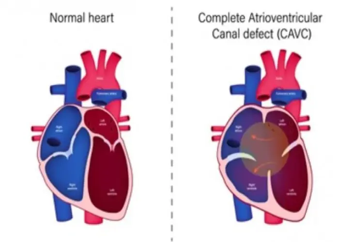
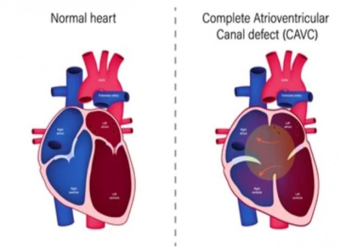
An atrioventricular (AV) canal defect is a type of congenital heart defect that affects the structure of the heart. It occurs when there is a hole in the wall (septum) separating the two upper chambers of the heart (the atria) and the two lower chambers of the heart (the ventricles). As a result, there is a mixing of oxygenated and deoxygenated blood, which can reduce the amount of oxygen that reaches the body.
AV canal defects can cause a variety of symptoms, including fatigue, shortness of breath, and rapid breathing, especially during physical activity. In severe cases, the heart may not be able to pump enough blood to meet the body's needs, which can lead to heart failure and other complications.
AV canal defects are typically diagnosed using a combination of medical history, physical examination, and imaging tests, such as an echocardiogram or a magnetic resonance imaging (MRI) scan. Treatment for AV canal defects may include medication to manage symptoms, surgery to close the hole in the septum, or a combination of both. In some cases, a catheter-based procedure may be recommended to correct the defect.
It is important to seek medical attention if you or your child has been diagnosed with an AV canal defect, as early and ongoing management can help prevent complications and improve overall heart function. Your healthcare provider can help determine the best course of treatment for your individual needs and monitor your progress over time.
Research Papers
Disease Signs and Symptoms
- Rapid breathing
- Swollen arms or hands
- Irregular heartbeats (arrhythmia)
- Rapid heartbeat (tachycardia)
- Excessive sweat
- Pale skin color (pallor)
- Poor growth or weight gain
- Loss of appetite
- Fatigue (Tiredness)
- Difficulty breathing (dyspnea)
- Swollen (Edema)
Disease Causes
Atrioventricular canal defect
Atrioventricular canal defect occurs before birth when a baby's heart is developing. Doctors aren't exactly sure what causes atrioventricular canal defect. Down syndrome might increase a person's risk of this congenital heart defect.
What happens in atrioventricular canal defect
Partial atrioventricular canal defect:
- A hole in the heart's wall (septum) separates the upper chambers (atria).
- Often the valve between the upper and lower left chambers (mitral valve) is abnormal and leaks blood (mitral valve regurgitation).
Complete atrioventricular canal defect:
- There's a large hole in the center of the heart where the walls between the upper chambers (atria) and the lower chambers (ventricles) meet. Oxygen-rich and oxygen-poor blood mix through that hole.
- Instead of separate valves on the right and left, there's one large valve between the upper and lower chambers.
- The abnormal valve leaks blood into the ventricles.
- The heart is forced to work harder and becomes larger.
Disease Prevents
Disease Treatments
Surgery is needed to repair a complete or partial atrioventricular canal defect. More than one surgery may be needed. Surgery to correct atrioventricular canal defect involves using one or two patches to close the hole in the heart wall. The patches stay in the heart permanently, becoming part of the heart's wall as the heart's lining grows over them.
Other surgeries depend on whether you have a partial or complete defect and what other heart problems you may have. For a partial atrioventricular canal defect, surgery to repair the mitral valve is needed so that the valve will close tightly. If repair isn't possible, the valve might need to be replaced.
For a complete atrioventricular canal defect, surgeons separate the abnormal large single valve between the upper and lower heart chambers into two valves. If separating the single valve isn't possible, heart valve replacement of both the tricuspid valve and the mitral valve might be needed.
After surgery
If the heart defect is repaired successfully, you or your child will likely have no activity restrictions.
You or your child will need lifelong follow-up care with a cardiologist trained in congenital heart disease. Your cardiologist will likely recommend a follow-up exam once a year or more frequently if problems, such as a leaky heart valve, remain.
Adults whose congenital heart defects were treated in childhood may need care from a cardiologist trained in adult congenital heart disease (adult congenital cardiologist) throughout life. Special attention and care may be needed around the time of any future surgical procedures, even those that do not involve the heart.
You or your child might also need to take preventive antibiotics before certain dental and other surgical procedures if either of you:
- Have remaining heart defects after surgery
- Received an artificial heart valve
- Received artificial (prosthetic) material during heart repair
- Will receive a heart valve prosthetic in the next six months
The antibiotics are used to prevent inflammation and infection of the lining of the heart (endocarditis).
Many people who have corrective surgery for atrioventricular canal defect don't need additional surgery. However, some complications, such as heart valve leaks, may require treatment.
Disease Diagnoses
Disease Allopathic Generics
Disease Ayurvedic Generics
Disease Homeopathic Generics
Disease yoga
Atrioventricular canal defect and Learn More about Diseases

Broken heart syndrome
Anal fissure
Cushing syndrome

Mesothelioma

Albinism
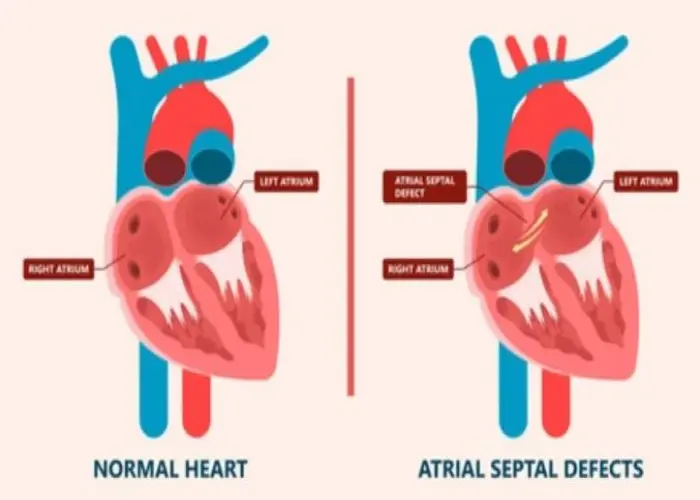
Atrial septal defect (ASD)

Heatstroke
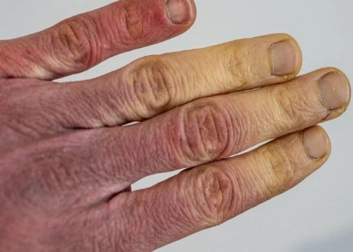
Raynaud's disease
Atrioventricular canal defect, AV block, AV node, অ্যাট্রিওভেন্ট্রিকুলার চ্যানেল ত্রুটি
To be happy, beautiful, healthy, wealthy, hale and long-lived stay with DM3S.
