 Welcome
“May all be happy, may all be healed, may all be at peace and may no one ever suffer."
Coarctation of the aorta

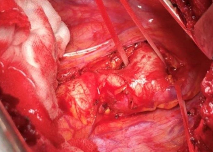
Coarctation of the aorta is a congenital heart defect in which the aorta, the main artery that carries oxygen-rich blood from the heart to the rest of the body, is narrowed or constricted. This narrowing can restrict blood flow to the body's organs and tissues, leading to a range of symptoms and complications.
The exact cause of the coarctation of the aorta is not known, but it is believed to be due to a combination of genetic and environmental factors. It is more common in males than females and may be associated with other congenital heart defects, such as a bicuspid aortic valve.
Symptoms of coarctation of the aorta may vary depending on the severity of the condition and may include high blood pressure, headaches, chest pain, leg cramps, shortness of breath, and fatigue. In severe cases, coarctation of the aorta can lead to heart failure, stroke, or even death.
Treatment for coarctation of the aorta typically involves surgery to repair or remove the narrowed segment of the aorta. In some cases, a balloon catheter may be used to widen the narrowed area of the aorta, a procedure known as balloon angioplasty. Long-term follow-up care is important to monitor for any recurrence of the narrowing, as well as to address any ongoing complications or associated health issues.
With appropriate treatment, the prognosis for coarctation of the aorta is generally good, and most people with the condition are able to lead normal, healthy lives. However, early diagnosis and intervention are important to prevent complications and improve outcomes.
Research Papers
Disease Signs and Symptoms
- Pale skin color (pallor)
- Fainting (syncope)
- Chest pain
- Nose bleeding
- Leg cramp
- Muscle weakness
- Headaches
- High blood pressure (hypertension)
- Difficulty breathing (dyspnea)
- Heavy sweating (diaphoresis)
- Irritability
- Shortness of breath (dyspnea)
Disease Causes
Coarctation of the aorta
Doctors aren't certain what causes coarctation of the aorta. The condition is generally present at birth (congenital). Congenital heart defects are the most common of all birth defects.
Rarely, coarctation of the aorta develops later in life. Conditions or events that can narrow the aorta and cause this condition include:
- Traumatic injury
- Severe hardening of the arteries (atherosclerosis)
- Inflamed arteries (Takayasu's arteritis)
Coarctation of the aorta usually occurs beyond the blood vessels that branch off to your upper body and before the blood vessels that lead to your lower body. This can often lead to high blood pressure in your arms but low blood pressure in your legs and ankles.
With coarctation of the aorta, the lower left heart chamber (left ventricle) of your heart works harder to pump blood through the narrowed aorta, and blood pressure increases in the left ventricle. This may cause the wall of the left ventricle to thicken (hypertrophy).
Disease Prevents
Coarctation of the aorta
Coarctation of the aorta can't be prevented, because it's usually present at birth. However, if you or your child has a condition that increases the risk of aortic coarctation, such as Turner syndrome, bicuspid aortic valve or another heart defect, or a family history of congenital heart disease, early detection can help. Discuss the risk of aortic coarctation with your doctor.
Disease Treatments
Treatment for coarctation of the aorta depends on your age at the time of diagnosis and the severity of your condition. Other heart defects might be repaired at the same time as aortic coarctation.
A doctor trained in congenital heart conditions will evaluate you and determine the most appropriate treatment for your condition.
Medication
Medication isn't used to repair coarctation of the aorta. However, your doctor may recommend it to control blood pressure before and after stent placement or surgery. Although repairing aortic coarctation improves blood pressure, many people still need to take blood pressure medication after a successful surgery or stenting.
Babies with severe coarctation of the aorta often are given a medication that keeps the ductus arteriosus open. This allows blood to flow around the constriction until the coarctation is repaired.
Surgery or other procedures
There are several surgeries to repair aortic coarctation. Your doctor can discuss with you which type is most likely to successfully repair your or your child's condition. The options include:
- Resection with end-to-end anastomosis. This method involves removing the narrowed segment of the aorta (resection) followed by connecting the two healthy sections of the aorta together (anastomosis).
- Subclavian flap aortoplasty. A part of the blood vessel that delivers blood to your left arm (left subclavian artery) might be used to expand the narrowed area of the aorta.
- Bypass graft repair.This technique involves bypassing the narrowed area by inserting a tube called a graft between the portions of the aorta.
- Patch aortoplasty. Your doctor might treat your coarctation by cutting across the narrowed area of the aorta and then attaching a patch of synthetic material to widen the blood vessel. Patch aortoplasty is useful if the coarctation involves a long segment of the aorta.
Balloon angioplasty and stenting
This procedure may be used as a first treatment for aortic coarctation instead of surgery, or it may be done if narrowing occurs again after coarctation surgery.
During balloon angioplasty, your doctor inserts a thin, flexible tube (catheter) into an artery in your groin and threads it through your blood vessels to your heart using X-ray imaging.
Your doctor places an uninflated balloon through the opening of the narrowed aorta. When the balloon is inflated, the aorta widens and blood flows more easily. Sometimes, a mesh-covered hollow tube (stent) is placed in the aorta to keep the narrowed part of the aorta open.
Disease Diagnoses
Disease Allopathic Generics
Disease Ayurvedic Generics
Disease Homeopathic Generics
Disease yoga
Coarctation of the aorta and Learn More about Diseases

Leprosy

Bradycardia

Colic

Pubic lice (crabs)
Polycystic kidney disease
Hemangioma
Gilbert's syndrome
Septic arthritis
Coarctation of the aorta, Coarctation, কোগটেইশন অব দি এওরটার
To be happy, beautiful, healthy, wealthy, hale and long-lived stay with DM3S.
