 Welcome
“May all be happy, may all be healed, may all be at peace and may no one ever suffer."
Retinal detachment
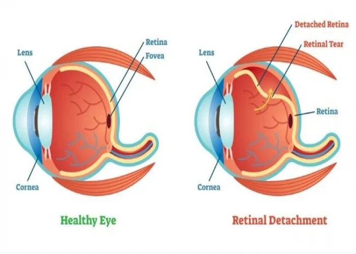

Retinal detachment is a serious eye condition that occurs when the retina, the thin layer of tissue at the back of the eye that sends visual signals to the brain, becomes detached from its normal position. This detachment can cause loss of vision and, if not treated promptly, can lead to permanent vision loss.
Retinal detachment can be caused by a variety of factors, including trauma, aging, myopia (nearsightedness), and certain medical conditions, such as diabetes. Symptoms of retinal detachment can include sudden flashes of light, the appearance of "floaters" or small spots in the visual field, and a shadow or "curtain" effect in the visual field.
Diagnosis of retinal detachment typically involves a comprehensive eye exam, including a dilated eye exam, in which the pupil is dilated with special eye drops to allow the healthcare professional to examine the retina more closely. In some cases, other tests, such as ultrasound imaging or optical coherence tomography (OCT), may be needed to evaluate the extent and location of the detachment.
Treatment for retinal detachment typically involves surgery, as the detachment will not reattach on its own. The specific type of surgery depends on the severity and location of the detachment but may involve a procedure to reattach the retina using lasers or cryotherapy, or a more invasive surgery such as a scleral buckle or vitrectomy.
It is important to seek medical attention promptly if you experience any symptoms of retinal detachment, as early diagnosis and treatment can help prevent permanent vision loss. Regular eye exams and early treatment of conditions such as diabetes or myopia can also help prevent retinal detachment from occurring.
Research Papers
Disease Signs and Symptoms
- Flashes of light in one or both eyes (photopsia)
- Blurred vision of eye
- A curtain-like shadow over your visual field
- Retinal detachment
Disease Causes
Retinal detachment
There are three different types of retinal detachment:
- Rhegmatogenous (reg-ma-TODGE-uh-nus). These types of retinal detachments are the most common. Rhegmatogenous detachments are caused by a hole or tear in the retina that allows fluid to pass through and collect underneath the retina, pulling the retina away from underlying tissues. The areas where the retina detaches lose their blood supply and stop working, causing you to lose vision.
- The most common cause of rhegmatogenous detachment is aging. As you age, the gel-like material that fills the inside of your eye, known as the vitreous (VIT-ree-us), may change in consistency and shrink or become more liquid. Normally, the vitreous separates from the surface of the retina without any complications — a common condition called posterior vitreous detachment (PVD). One complication of this separation is a tear.
- As the vitreous separates or peels off the retina, it may tug on the retina with enough force to create a retinal tear. Left untreated, the liquid vitreous can pass through the tear into the space behind the retina, causing the retina to become detached.
- Tractional. This type of detachment can occur when scar tissue grows on the retina's surface, causing the retina to pull away from the back of the eye. Tractional detachment is typically seen in people who have poorly controlled diabetes or other conditions.
- Exudative. In this type of detachment, fluid accumulates beneath the retina, but there are no holes or tears in the retina. Exudative detachment can be caused by age-related macular degeneration, injury to the eye, tumors or inflammatory disorders.
Disease Prevents
Disease Treatments
Surgery is almost always used to repair a retinal tear, hole or detachment. Various techniques are available. Ask your ophthalmologist about the risks and benefits of your treatment options. Together you can determine what procedure or combination of procedures is best for you.
Retinal tears
When a retinal tear or hole hasn't yet progressed to detachment, your eye surgeon may suggest one of the following procedures to prevent retinal detachment and preserve vision.
- Laser surgery (photocoagulation). The surgeon directs a laser beam into the eye through the pupil. The laser makes burns around the retinal tear, creating scarring that usually "welds" the retina to underlying tissue.
- Freezing (cryopexy). After giving you a local anesthetic to numb your eye, the surgeon applies a freezing probe to the outer surface of the eye directly over the tear. The freezing causes a scar that helps secure the retina to the eye wall.
Both of these procedures are done on an outpatient basis. After your procedure, you'll likely be advised to avoid activities that might jar the eyes — such as running — for a couple of weeks or so.
Retinal detachment
If your retina has detached, you'll need surgery to repair it, preferably within days of a diagnosis. The type of surgery your surgeon recommends will depend on several factors, including how severe the detachment is.
- Injecting air or gas into your eye. In this procedure, called pneumatic retinopexy (RET-ih-no-pek-see), the surgeon injects a bubble of air or gas into the center part of the eye (the vitreous cavity). If positioned properly, the bubble pushes the area of the retina containing the hole or holes against the wall of the eye, stopping the flow of fluid into the space behind the retina. Your doctor also uses cryopexy during the procedure to repair the retinal break.
- Fluid that had collected under the retina is absorbed by itself, and the retina can then adhere to the wall of your eye. You may need to hold your head in a certain position for up to several days to keep the bubble in the proper position. The bubble eventually will reabsorb on its own.
- Indenting the surface of your eye. This procedure, called scleral (SKLAIR-ul) buckling, involves the surgeon sewing (suturing) a piece of silicone material to the white of your eye (sclera) over the affected area. This procedure indents the wall of the eye and relieves some of the force caused by the vitreous tugging on the retina.
- If you have several tears or holes or an extensive detachment, your surgeon may create a scleral buckle that encircles your entire eye like a belt. The buckle is placed in a way that doesn't block your vision, and it usually remains in place permanently.
- Draining and replacing the fluid in the eye. In this procedure, called vitrectomy (vih-TREK-tuh-me), the surgeon removes the vitreous along with any tissue that is tugging on the retina. Air, gas or silicone oil is then injected into the vitreous space to help flatten the retina.
- Eventually the air, gas or liquid will be absorbed, and the vitreous space will refill with body fluid. If silicone oil was used, it may be surgically removed months later.
- Vitrectomy may be combined with a scleral buckling procedure.
After surgery your vision may take several months to improve. You may need a second surgery for successful treatment. Some people never recover all of their lost vision.
Disease Diagnoses
Disease Allopathic Generics
Disease Ayurvedic Generics
Disease Homeopathic Generics
Disease yoga
Retinal detachment and Learn More about Diseases

Heart murmurs

Pregnancy Abodominal Pain
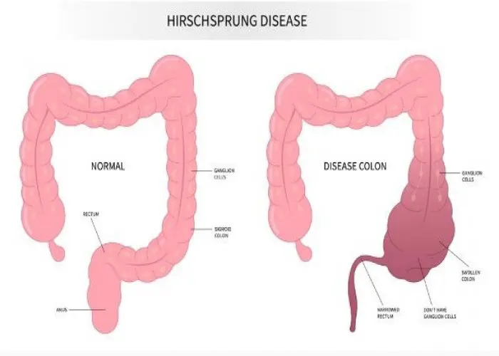
Hirschsprung's disease

MCAD deficiency

Uterine prolapse

CSF leak (Cerebrospinal fluid leak)
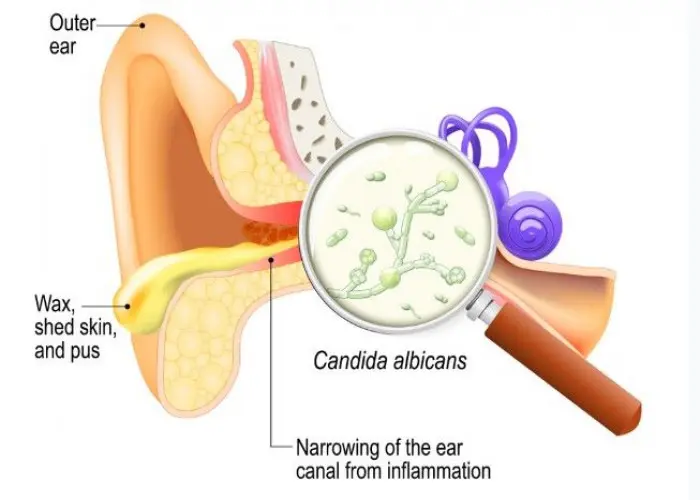
Swimmer's ear

Kidney stones
retinal detachment, রেটিনার ডিটাসমেন্ট
To be happy, beautiful, healthy, wealthy, hale and long-lived stay with DM3S.